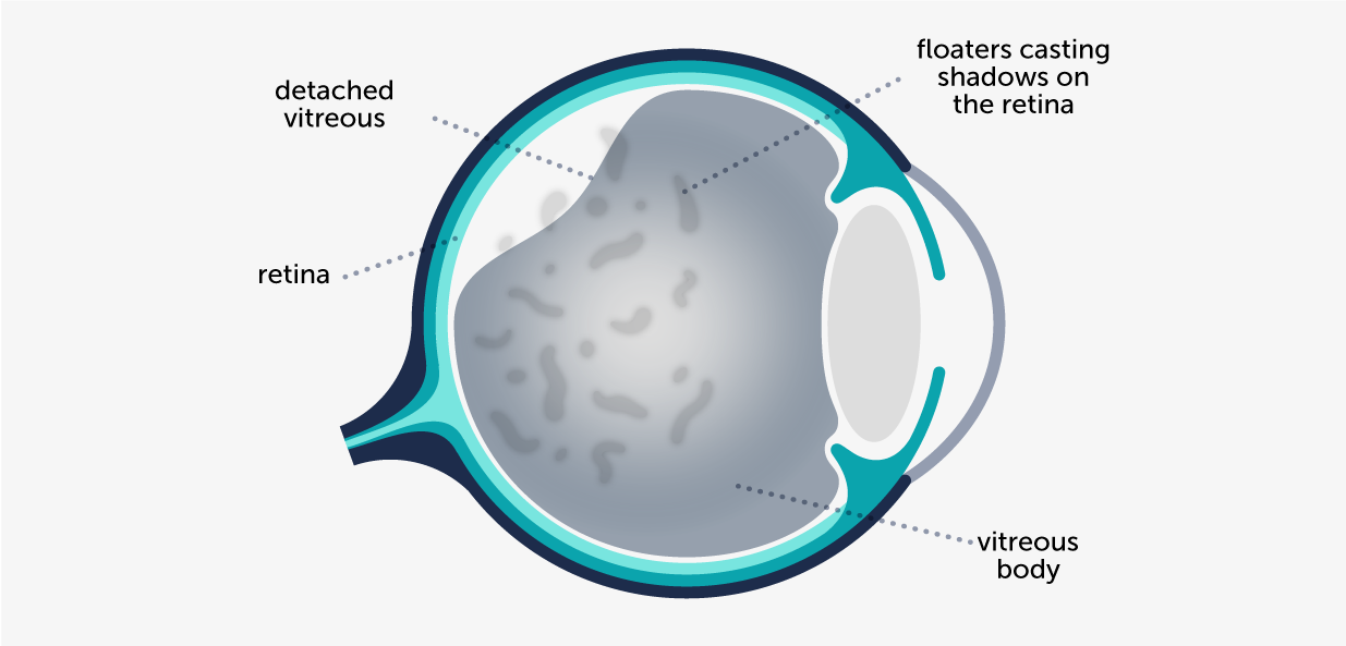
Floaters should not be an issue of concern, but vitreous detachment should be urgently examined. Advanced age, myopia, cataract surgery and eye traumas are the main risk factors for a vitreous detachment.
Small translucent spots or shadows
They appear in isolation
Spots are high in number
They have the shape of a spider web
Possible flashing lights
Possible vision decrease
| Symptoms of eye floaters | Symptoms of a vitreous detachment |
|---|---|
| It is not a serious condition | It requires an urgent examination to rule out more serious problems, such as retinal tears or a retinal detachment. |
The so-called floaters (vitreous syneresis in medical terms) are mobile spots that appear on the visual field and move with eye movement.
They appear as small translucent spots or shadows, and are better perceived with light backgrounds, such as a white wall, the sky or a computer screen.
It is a natural ageing process.
Inside the eye, we have the vitreous cavity, which fills most of the eyeball. The inner walls of such cavity are the retina, and they contain a jelly-like material called vitreous humour, which is glued on the retina by multiple anchoring points.
Floaters appear as vitreous humour naturally deteriorates over time. Consequently, some areas liquefy and some others are condensed in lumps or threads, which is what we see as floaters or shadows.
A vitreous detachment is the separation of this jelly-like substance the fills the vitreous cavity from its attachment points on the retina. This causes a sudden appearance of spots in the form of spider webs and, in some cases, the vision of flashing lights.
The vitreous detachment isn’t a serious condition by itself, but when it is detached, it pulls from the retina, and it may cause a retinal hole or tear.
A retinal tear needs to be urgently treated with laser at the consulting room. In case it is not treated on time, it may lead to a retinal detachment with more severe consequences to vision.
In the case of floaters, the patient reports seeing small translucent spots sometimes. They are think and not high in number, and they move with eye movements. Usually patients have been experiencing it for a while and they have not observed any change in its form or pattern.
Conversely, in posterior vitreous detachment, floaters appear suddenly, in high number, as black spots floating in the visual field. Patients describe it as a spider web or a net in front of their visual field, which moves in accordance to eye movements.
Sometimes it is also accompanied by a certain decrease in vision.
Some patients also report seeing sparkles in the corner of their visual field, as well as some flashes called photopsias. There are light stimulations that may last for a second, and are mostly seen at night or in dim light conditions. They usually appear on a corner of the visual field, and have a linear or semi-circular pattern.

Vision of small translucent floaters detected some time ago and with no changes in pattern is not an urgent nor serious condition. Scheduling an ophthalmology examination is nonetheless recommended.
But whenever the floaters appear in a sudden way and in high numbers, creating a spider web or a spot net, it is important to urgently be examined. In such cases, it is important to rule out more serious conditions, such as retinal tears or a retinal detachment.
Main risk factors of posterior vitreous detachment posterior are:
It is important that people who have suffered a vitreous detachment are vigilant, as symptoms may appear in the other eye, because the risk of suffering this condition is higher in these patients.
Usually, the patient gets used to the situation after some months of the onset of the spots. With the mitigation of the spot opacity, and due to a visual adaptation process of the patient, the spot perception is reduced, and in some cases, it may even disappear.
Even though they are quite annoying, at ICR we do not recommended to perform a surgical intervention for floaters or retinal detachment. We consider it does not counterbalance the eye surgery risks.
In any case, a good ophthalmic monitoring is important to prevent more serious conditions.
In very specific cases in which vitreous condensations are important and patients are highly uncomfortable due to the effect they have on their visual quality, a surgery may be considered. The surgical technique used is called vitrectomy, and it consists in eliminating all the jelly-like fluid from within the eye, which is the cause of floaters, and replace it by another substance.
It is important to bear in mind that, like in any other surgery, vitrectomy may have complications:

Contact us or request an appointment with our medical team.