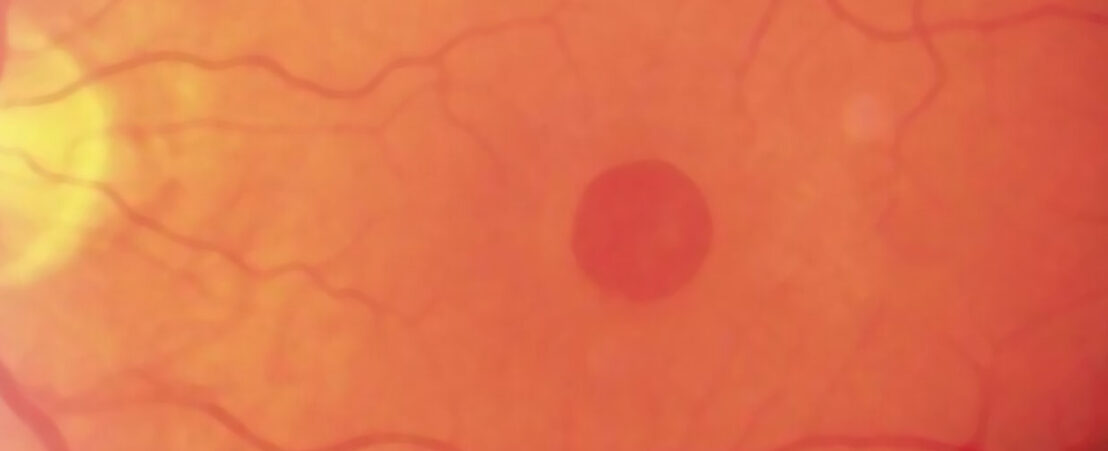
Macular hole

Medical content revised by - Last revision 31/08/2020
Macular hole

Symptoms

A macular hole causes loss of central vision and image distortion in the eye. The distortion is manifested as a bending or waviness of straight lines or objects can begin to look bent or wavy and numbers and letters jump out of line.

Other symptoms manifested by patients are a central grey spot or a blind spot in the central vision and difficulty reading.

The macular hole usually manifests itself in an acute way overnight, but sometimes the visual loss is more gradual and takes place during weeks or months. Sometimes patients don’t realize it until they come to cover the eye that is not affected.


Typical signs

In the early stages of the disease, a yellow injury in the fovea is detected. The fovea is the part of the retina located in the centre of the macula and is responsible for the detailed vision, necessary for reading.

When the hole is consolidated, it is displayed in the fovea.


Types of macular hole

  • Grade 1: A detachment of the foveal area of the retina without full retinal hole is detected.
  • Grade 2: A small full retinal hole is detected in the foveal area.
  • Grade 3: There’s an increase in size of the hole (of 400 microns)
  • Grade 4: The hole is associated with a posterior vitreous detachment. The vitreous is the jelly-like substance that fills the inside of the eye

Causes

IDIOPATHIC

It appears in healthy patients without evidence of other eye diseases. It is the most common type of macular hole and is related to advanced age.

SECONDARY

  • Myopia: Patients with myopia magna (greater than 9 dioptres) have an increased risk of macular hole due the anatomical features of the myopic eye.
  • Eye trauma.
  • Chronic inflammation of the macula: In cases of chronic cystoid macular oedema, it is secondary to an eye inflammation, previous intraocular surgery, vascular disorders of the retina…
  • Intraocular surgery.

Management of the patient with macular hole

Determine whether any of the above-mentioned causes is responsible for the macular hole. If that’s the case, both problems would need to be treated.

Before solving the macular hole, we need to assess the degree of cataract of the patient. In patients who also suffer from a cataract, a combined surgery consisting in cataract extraction and macular hole surgery is recommended. This is performed in an only procedure in order to avoid the need to perform a second intervention later in time.

  • Personal history: drugs, trauma, systemic diseases and eye diseases.
  • Full eye examination, including:
    • Distant and near vision, pupil examination and refraction.
    • A biomicroscopy under pupil dilation, with direct lighting and backlighting, in order to study the cornea, the anterior chamber, and the existence of a cataract, as well as to rule out any ocular inflammation.
    • A tonometry to determine eye pressure.
    • A macular study of the retina, paying special attention to the macula in order to determine the extent of the hole. It is also very important to rule out vitreous inflammation and examine the retina in depth.
    • A macular OCT to confirm the diagnosis, determine the size and shape of the macular hole, rule out a cystoid macular oedema and assess the status of the different retinal layers.
    • A fluorescein angiography. In some patients, it may be necessary to complete the study with this test in order to rule out other retinal diseases.
    • Ultrasound or interferometry biometry: when a cataract surgery is considered, keratometric data becomes necessary and the measurement of the axis by means of type A ultrasound or laser is used to calculate the power of the intraocular lens that the patient will need.
    • The assessment of corneal endothelium is also necessary, usually by means of a slit lamp, and sometimes with endothelial cell count.

Treatment

Macular hole surgery is performed in order to:

  • improve the vision and reduce the image distortion in patients with symptomatic visual disturbances.
  • avoid the progressive visual loss that occurs over time.

Cataract extraction is performed in order to:

  • improve the view of the patient.
  • avoid the need for a second intervention due to the progression of the cataract later in time, which is something very common in patients with some degree of cataract and who are over 60 years old.

The cataract extraction allows us to correct any refractive errors that the patient may have.


Monitoring

Patients with stage 2, 3 and 4 macular holes generally require surgery.

Patients with early stage macular holes (stage 1) that do not require surgery should be examined from time to time to assess whether there has been a disease progression and they will have to perform a self-monitoring at home with Amsler grid in order to determine whether there are changes in the distortion.

Patients should go to see the ophthalmologist before the scheduled date if there is a symptomatic decrease in vision or an increase in distortion.

Each examination will consist in the assessment of any vision loss or increase in image distortion. A macular OCT will be performed in order to assess whether there has been a progression in the macular hole stage.

Related articles

Age-Related Macular Degeneration (ARMD)

Age-related macular degeneration or AMD is a disease that affects the central part of the retina, the macula, and, as its name indicates, it is a pathology that progresses as the patient’s age increases. AMD causes central vision problems and can lead to blindness if it is not treated early. Types of AMD Currently, we […]

Macular epiretinal membrane

Symptoms Macular epiretinal membrane causes a slow and progressive visual loss lasting months or years, and affecting one or both eyes. Generally, it causes image distortion, and undulation of straight lines. Numbers and letters can appear to be in another line, and perception of colors may be decreased. Signs The typical sign is a  semitransparent […]

Vitreous detachment

What is Vitreous detachment? If floaters are small, translucent and isolated, and provided they always have the same shape and pattern, there is nothing to worry about. Scheduling an ophthalmology examination is nonetheless recommended. If floaters appear abruptly and in a great number, sometimes in a manner described as a spider web that blocks part […]

Cataract surgery

At ICR we work with enthusiasm, involvement and commitment to ensure that our patients recover their vision and that they do so depending as little as possible on their glasses. Our cataract specialists will advise each patient the best option based on their needs, lifestyle and medical history.

Do you have any questions?

Contact us or request an appointment with one of our Retina and Vitreous specialists.

Visit us at:

ICR Ganduxer Headquarters

C/ Ganduxer,117
08022 Barcelona See map

ICR Pau Alcover

C/ Pau Alcover 67
08017 Barcelona See map

ICR Cerignola

C/ Cerignola, 14
08022 Barcelona See map

ICR Terrassa

C/ Del Nord, 77
08221 Terrassa See map

ICR Service at Clínica Ntra. Sra. del Remei

C/ de l'Escorial,148
08024 Barcelona See map

Clínica Bonanova Surgical Center

Passeig Bonanova, 22
08022 Barcelona See map

Clinica Bonanova Tests and Treatments

C/ Mandri, 63
08022 Barcelona See map

ICR Ganduxer Headquarters

Telephone number +34 93 378 23 19
Emergencies phone number: +34 93 418 72 27

Opening hours:
Monday to Friday 8 a.m. to 9 p.m.
Saturdays 9 a.m. to 2 p.m.

How to get there More information

ICR Pau Alcover

Telephone number (+34) 93 378 23 19

Opening hours:
Monday to Friday from 8 am to 9 pm. Saturday from 8 am to 2 pm.

How to get there More information

ICR Cerignola

Telephone number +34 93 378 23 19

Opening hours:
Monday to Friday 8 a.m. to 9 p.m.

How to get there More information

ICR Terrassa

Telephone number +34 93 736 34 40

Opening hours:
Monday to Friday from 8:30 a.m. to 2 p.m. and 3 p.m. to 8 p.m.

How to get there More information

ICR Service at Clínica Ntra. Sra. del Remei

Telephone number 902 10 10 50

Opening hours:
Monday to Thursday from 3 p.m. to 8 p.m. and Monday mornings from 8 a.m. to 2 p.m.

How to get there More information

Clínica Bonanova Surgical Center

Telephone number 934 34 09 25

Opening hours:
Monday to Friday from 7:30 a.m. to 8:30 p.m.

How to get there More information

Clinica Bonanova Tests and Treatments

Telephone number +34 93 434 27 70

How to get there More information