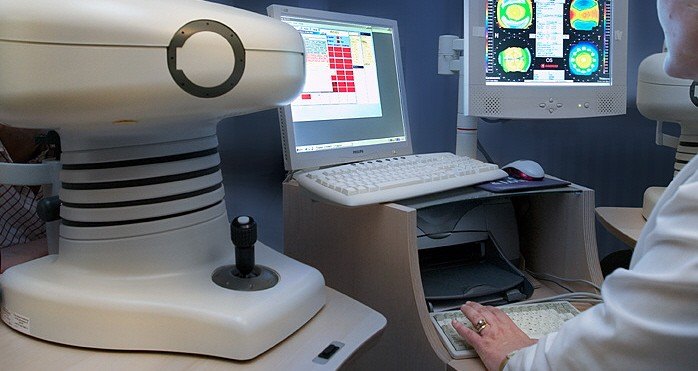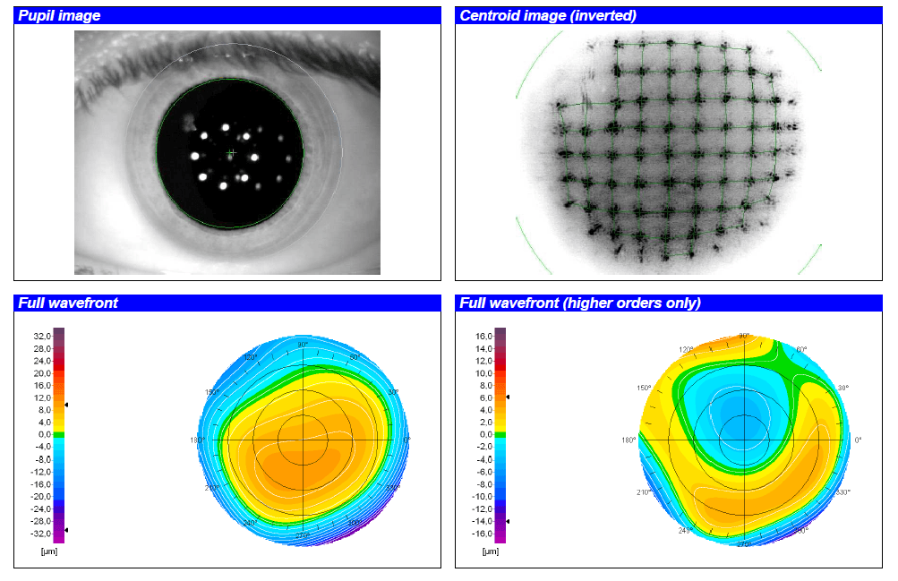
Aberrometry is a non-invasive ophthalmological test that allows studying the optical quality of the visual system. By analyzing the wavefront, structures involved in the refraction of light, such as the cornea (the outer, transparent layer through which light penetrates the eye) and the crystalline lens (the inner lens), are studied. Through this test, the ophthalmologist will be able to obtain information about the quality of the images perceived by the patient.
It is used to guide diagnoses, as well as to evaluate and follow up patients interested in refractive surgery (analysis of the prescription, measurement of pupil size, etc.) both in the preoperative and postoperative studies, in order to personalize the treatment as much as possible.
Therefore, aberrometry is not used to detect a particular pathology, but rather to analyze the structures of the eye that may be affecting the quality of the patient’s vision. Irregularities in the shape and transparency of the tissues, as well as optical defects, are elements that intervene in the result of an aberrometry and cannot always be corrected with glasses or contact lenses.
No particular preparation or pupil dilation is required (unless specifically indicated), but it is necessary to perform the test without contact lenses. Therefore, it is recommended to stop using contact lenses at least the day before the test.
Aberrometry is a non-invasive technique, so it does not require any direct contact with the eye and does not produce any adverse reactions. After the test, therefore, any day-to-day activity can be performed without any problem.
The test requires an aberrometer, a measuring instrument also known as a wavefront analyzer.
The aberrometer measures the deformation of laser beams, of very low power, which are projected into the eye. These are reflected off the back of the eye and returned to the measuring device. If the emitted beams are identical to those received, the eye is optically perfect. If not, specialists call this an optical aberration.
The test is performed in a seated position and does not require eye contact. With the chin placed on a chin rest and the forehead well placed forward, the patient must fix the eyes on a central luminous pattern inside the lens without moving the eyes or the head.
This test takes only a few seconds and is performed by an optometrist. Once the data has been collected, the ophthalmologist will be in charge of interpreting the results and transmitting them during the next visit.

No, since it is a non-invasive technique that does not contact the eye, it does not cause any type of discomfort.
No, this test does not require you to be accompanied by another person. In some exceptional cases, it is requested to perform it under pupillary dilation. In this case, your vision will be blurred by the eye drops and you will not be able to drive.
It is not necessary to fast for the test to be performed.
The data is obtained at the same time the test is performed. If a report is required by an ophthalmologist, the test report will be sent to you within a few days.
The optometrist is the one in charge of performing the test and has the knowledge to confirm its correct performance. The one who must interpret and report the results obtained is the ophthalmologist, who will do so taking into account the clinical context after a complete anamnesis and examination of the patient.
Yes, after the test you can perform any daily activity, such as showering or driving, except if you have had pupillary dilation.
Yes, there is no inconvenience in using make-up after this test.

Contact us or request an appointment with our medical team.