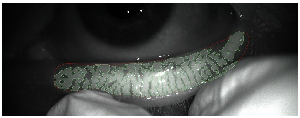
Meibography allows to evaluate the morphology of the Meibomian glands, located in the lower and upper eyelids, and to determine whether they fulfill their function of providing the necessary fats to limit tear evaporation.
It is a non-invasive technology that allows:
Meibography is performed to detect possible Meibomian gland dysfunction (MGD) and to monitor their correct functioning, since they are responsible for producing the meibomium, a complex lipid layer that forms part of the tear film and whose function is to ensure the stability of the tear, prevent its evaporation and thus protect the surface of the eye.
It is essential to check that the Meibomian glands are not obstructed to avoid complications such as dry eye syndrome or to know, through meibography, the degree of involvement and to be able to guide, if possible, the treatment of dry eye with pulsed light (IPL) to stimulate the functioning of these glands.
Meibomian gland dysfunction, also called posterior blepharitis, is the main cause of evaporative dry eye.
In addition, the tear film has great physiological and optical importance for the correct functioning of the human eye, so the diagnosis of any type of alteration, whether in its secretion mechanisms, stability or quality, of one or all of its layers is of great diagnostic help in visual health.
The test does not require pupil dilation, but it is necessary to come on the day of the test without having applied cream and/or make-up on the face and without contact lenses in order to obtain the results properly.
Meibography is a non-invasive technique, so it does not require any direct contact with the eye and does not produce any adverse reactions. After the test, therefore, any day-to-day activity can be performed without any problem.
This test takes only a few seconds and is performed by an optometrist. Once the data has been collected, the ophthalmologist will be in charge of interpreting the results and transmitting them during the next visit.
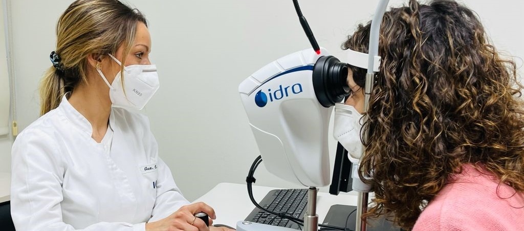
Meibography is performed monocularly, first one eye and then the other. The examination is performed seated and does not require contact with the eye surface. With the chin placed on a chin rest and the forehead well forward, the patient should fix the gaze towards a central point of the equipment. Throughout the test, the patient should follow the professional’s instructions.
In order to obtain images of the Meibomian glands of both the upper and lower eyelids, it will be necessary to apply light pressure on the outer palpebral area.
This test takes a few minutes and is performed by specialized staff. Once the information has been collected, the ophthalmologist will be in charge of interpreting the results and transmitting them during the next visit.

This is an infrared system that allows images to be obtained non-invasively and without coming into contact with the eye. However, the pressure exerted on the outer eyelid area in order to obtain images of the glands may cause slight discomfort.
No, this test does not require you to be accompanied by another person.
It is not necessary to fast for the test to be performed.
The data is obtained at the same time the test is performed. If a report is required by an ophthalmologist, the test report will be sent to you within a few days.
The technician is the one in charge of performing the test and has the knowledge to confirm its correct performance. The one who must interpret and report the results obtained is the ophthalmologist, who will do so taking into account the clinical context after a complete anamnesis and examination of the patient.
Yes, after the test you can perform any day-to-day activity, such as showering or driving.

Contact us or request an appointment with our medical team.