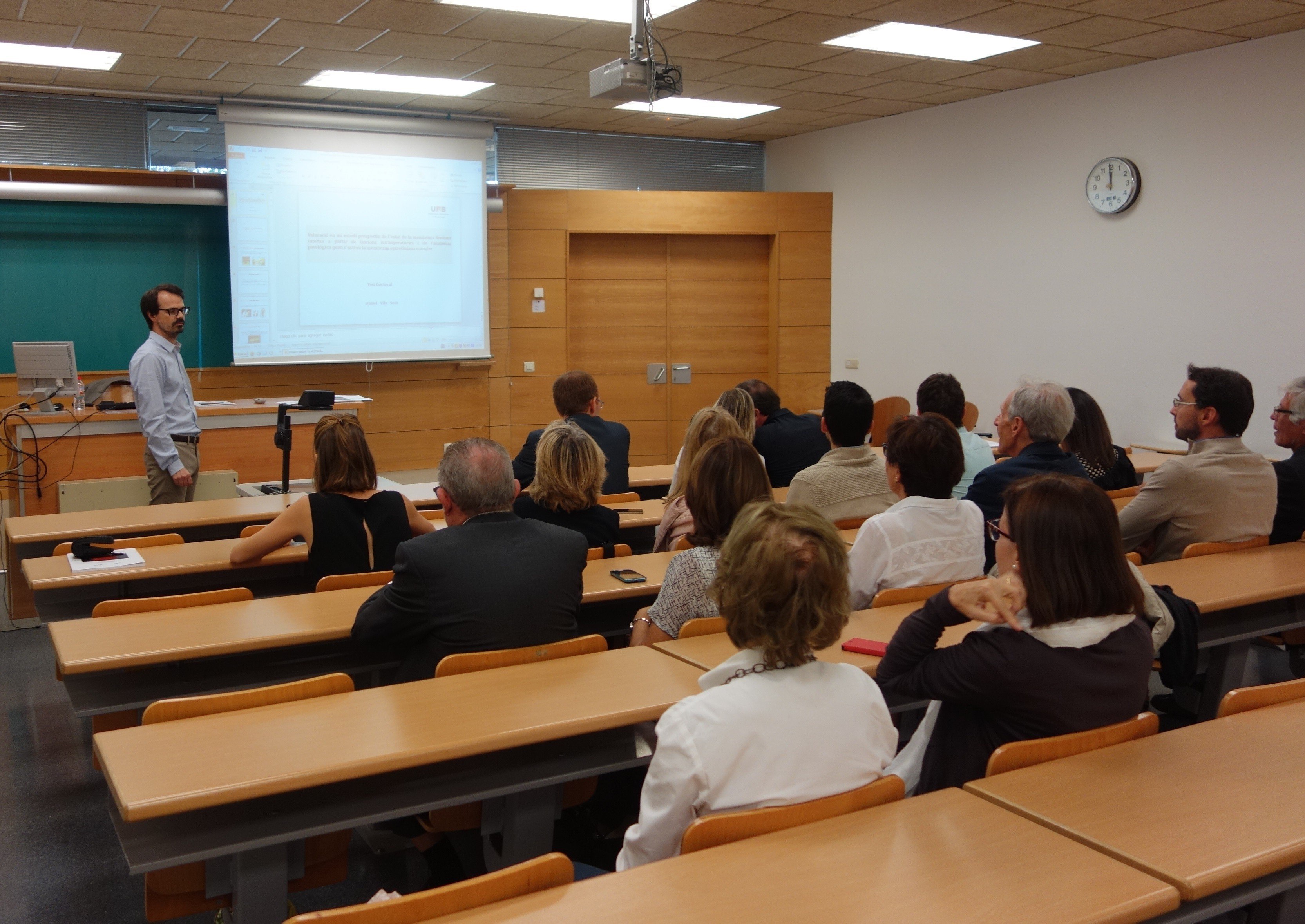
On 1st October, Dr. Daniel Vila presented his doctoral thesis defence at Universitat Autònoma de Barcelona under the title Assessment in a prospective study of the inner limiting membrane state from intra-operative staining and pathologic anatomy upon removal of macular epiretinal membrane, codirected by ICR Medical Director, Dr. Ignasi Jürgens.
His doctoral thesis had the aim of analysing the state of the inner limiting membrane (ILM) upon removal of macular epiretinal membrane (MEM) and determine the rate of concordance between assumedly removed tissues using intraoperative staining and the results of the samples pathological anatomy.
In order to achieve his goal, Dr. Vila carried out a prospective study in which patients diagnosed with idiopathic MEM were selected. Those patients were distributed randomly. In one group he sought to extract only the MEM (group M) and in another sequentially both MEM and ILM (group L). All patients underwent a pars plana vitrectomy and MEM removal with trypan blue staining. Afterwards, the state of ILM was intraoperatively assessed under brilliant blue G. In all patients from L group, ILM was removed and all obtained samples were anatomopathologically analyzed with an optical microscope.
The study was carried out in 26 patients from an average age of 70,65 years, divided in two groups: group M, counting 11 subjects, and group L, counting 15. The average monitoring time was 15,35 months and ILM removal patterns were as follow. In group M, removal was complete in 8 of the 11 patients and partial in 3 of the 11 patients. In group L, the removal was in block in 9 of the 15 patients, partial in 5 of the 15 patients and sequential in 1 of the 15 patients. In only 3,8 % of the total patients, IML remained unaffected after MEM removal.
Dr. Vila processed 32 surgical samples containing either MEM and ILM, either just MEM, either just ILM. A high rate of concordance of 84,37 % was detected between tissues assumedly removed through intraoperative staining. As for the samples assumedly containing IML, the rate of concordance reached 96,7 %.
The study allowed Dr. Vila to determine that it is technically difficult to remove MEM in isolation and that there is a high rate of concordance between tissues assumedly removed using operative staining and the results of pathological anatomy of samples, which is higher for ILM than for MEM.

Contact us or request an appointment with our medical team.