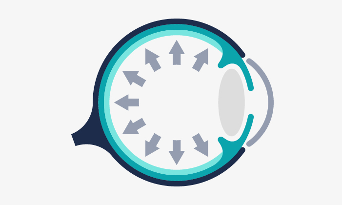
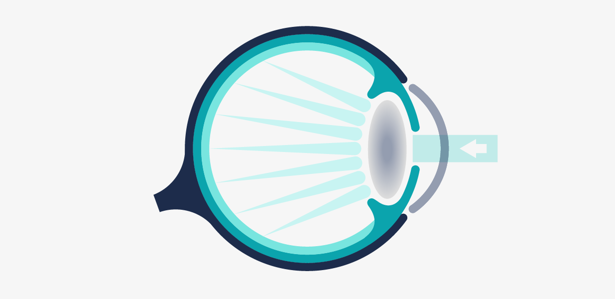
Ocular and orbital ecography, also known as ultrasonography or ocular ultrasound, is an imaging test that allows us to observe the structures of the eyeball even when there is a lack of transparency or opacity of the cornea or when we want to observe in detail parts of the orbit and the tissues around the eye.
There are several types of ocular ultrasound and each one is suitable for specific cases:
It is used to:
Through this test, images and videos of the inside of the eye and the orbit can be obtained. It is used to:
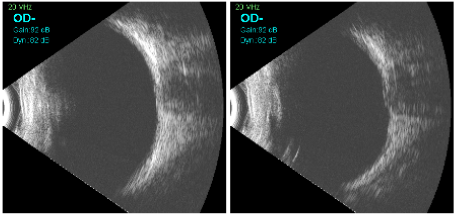
It is a high frequency ultrasound and is used to obtain images of the anterior segment of the eyeball. Obtains high definition images that are necessary to assess before implantation of phakic lenses in refractive surgery and patients with:
Ultrasounds A and B do not require any particular preparation or pupil dilation. For these tests it is not necessary to take a break from contact lenses, but it is advisable not to wear them.
However, the UBM should be performed without contact lenses and anesthetic drops are administered for the placement of a separator between the eyelids, thus avoiding the possible discomfort of the separator.
Ultrasounds A and B are considered a non-invasive technique, so they do not require direct contact with the eye and do not produce adverse reactions. After the test, therefore, any day-to-day activity can be performed without any problem.
As for UBM, when administering anesthetic drops, the patient will not be able to use contact lenses until a couple of hours after the drops have been administered.
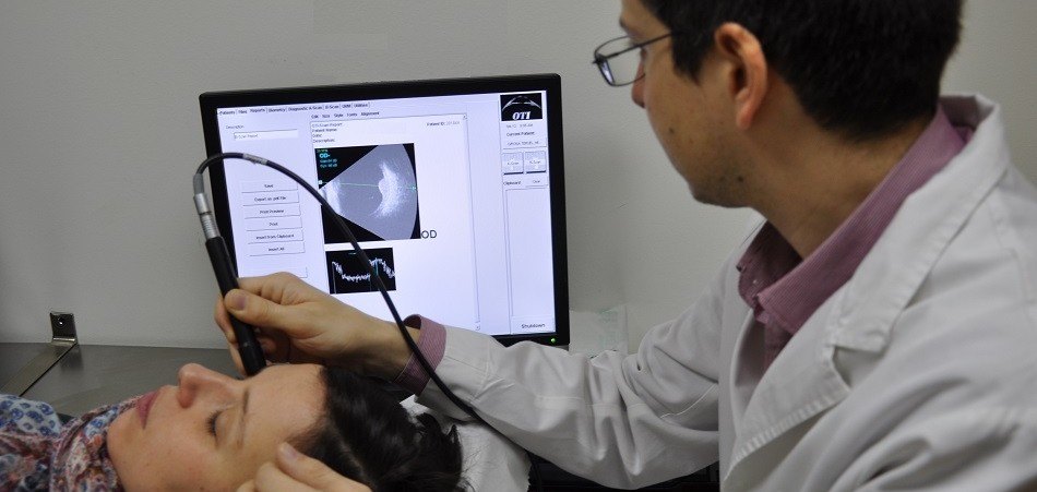
For ultrasounds A and B, a small amount of gel is applied over the ultrasound probe and the probe is placed gently against the eyelids to perform the examination. The patient should remain with the eyes closed while the optometrist performs the test.
In the case of UBM, anesthetic drops are administered in order to facilitate keeping the eyes open through a separator, the ocular surface is covered with saline to ensure transmission of the ultrasound signal, and a probe is gently slid over the saline, which never comes into contact with the eye.
The test takes only a few minutes and is performed by an ophthalmologist. Once the data has been collected, the ophthalmologist will be in charge of interpreting the results and transmitting them during the next visit.
Ultrasounds A and B are considered a non-invasive technique, they do not contact the eye and therefore do not cause any type of discomfort.
In the case of UBM, it is slightly invasive and therefore the discomfort is minimal.
No, this test does not require you to be accompanied by another person.
No, it is not necessary to fast for the test.
The results are obtained at the same time the test is performed. If a report is required by an ophthalmologist, the test report will be sent to you within a few days.
The optometrist is the one in charge of performing the test and has the knowledge to confirm its correct performance. It is up to the ophthalmologist to interpret and report the results obtained, who will do so in the clinical context after a complete anamnesis and examination of the patient.
Yes, after the test it is possible to carry out any daily activity, such as driving or showering.
Yes, there is no inconvenience in using make-up after this test.

Glaucoma entails a loss of the vision field, usually asymptomatic until later stages of the disease. If it is not treated properly, it can induce blindness.
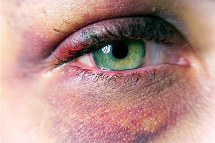
There are different kinds of eye injuries, caused by blunt or perforating objects as well as chemical products. If not treated properly, they could have serious consequences.
Our goal is to ensure that our patients recover their vision and that they do so depending as little as possible on their glasses.
Contact us or request an appointment with our medical team.


 Request an appointment