Specular microscopy or Endothelial cell counting

Medical content revised by - Last revision 27/01/2022
Specular microscopy or Endothelial cell counting

What is Specular Microscopy?

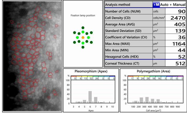
Specular microscopy is an optical test used to study the cornea, the transparent cellular layer that covers the eye, specifically the structure known as the endothelial tissue of the cornea, as well as to determine the number, shape and size of its endothelial cells. By means of the endothelial count, high-resolution images are obtained, so that an analysis of these images can be performed, a morphometric study of the cells can be made and possible lesions in this structure can be detected.

In general, a young healthy person shows in this internal layer a uniform pattern of hexagonal cells with the same size. This pattern can be altered as we grow up, due to pathological causes or surgical procedures. As a consequence of this change, there may be a loss of cells and even in situations of advanced cell loss, a decrease in vision.


What is it used for

Specular microscopy is mainly used for the systematic preoperative examination of patients who want to have cataract surgery or phakic lens implantation surgery. It is also used for the diagnosis and follow-up of corneal degeneration and dystrophies.


Before the test

The technique does not require any prior preparation, although it is important to perform it without the lenses in order to obtain adequate results.

In case of suspicion of altered results due to the use of contact lenses, it is recommended to repeat the test after 48 hours of rest.


After the test

Specular microscopy is a non-invasive technique, so it does not require direct contact with the eye and does not cause adverse reactions. After the test, therefore, any day-to-day activity can be carried out without any problem.


How is it done

Aquesta tècnica es basa en projectar un feix de llum amb un determinat angle sobre la còrnia de l’ull. La llum es reflecteix a l’endoteli corneal i és recollida altra vegada per l’instrument, que amplifica la zona il·luminada i analitza el patró cel·lular, determinant la densitat de cèl·lules a la zona de la captura. Una vegada recollides les dades, l’oftalmòleg serà l’encarregat d’interpretar els resultats i transmetre’ls durant la pròxima visita.

El departament encarregat de realitzar la prova és el Departament d’Optometria, que és qui realitza totes les proves oftalmològiques necessàries per al diagnòstic i el seguiment de les diferents patologies oculars i de tot el sistema visual. El Departament compta amb els equips diagnòstics més avançats i amb la tecnologia més moderna, i els seus prop de 50 optometristes estan formats específicament per a la realització d’aquestes proves.

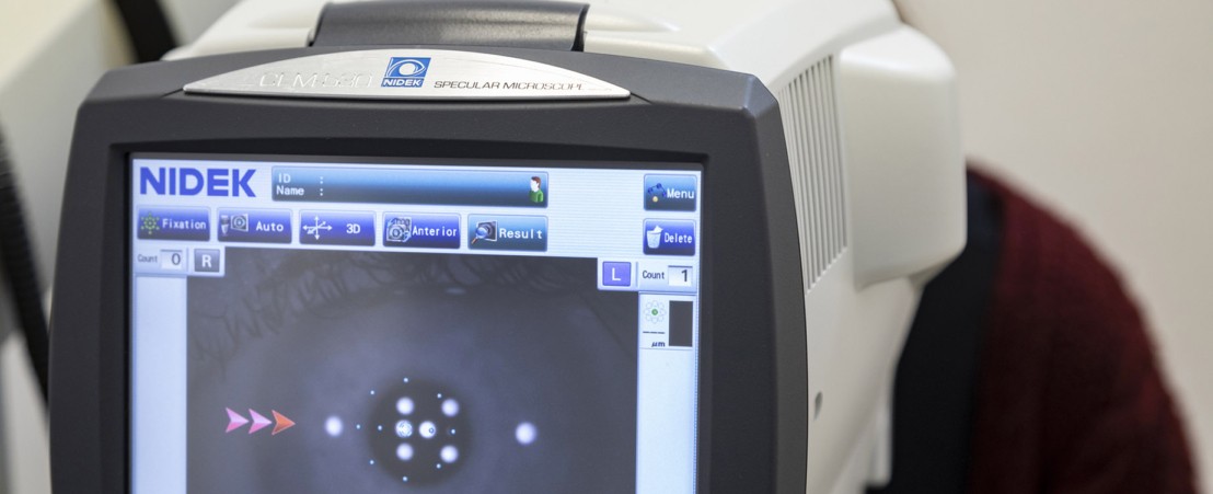
The test is performed using a non-contact specular microscope, which evaluates the cells of the most internal corneal layer, studies their density, size and shape. The microscope used is the CEM-530, the equipment besides characterizing the endothelial layer allows to know the value of the corneal thickness as a pachymeter.

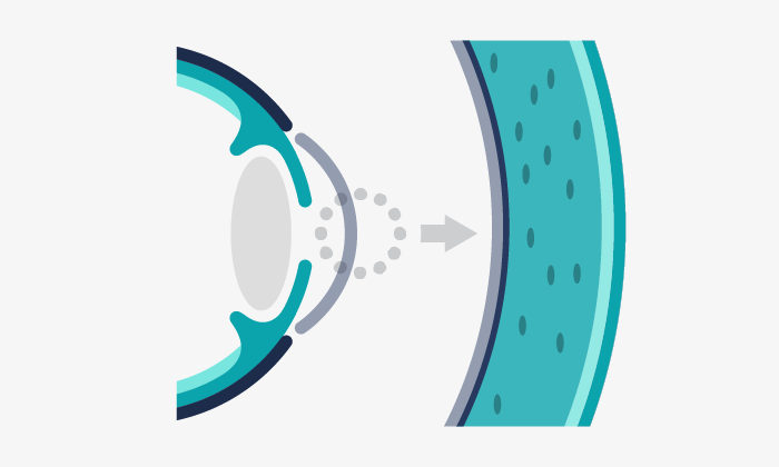
This technique is based on projecting a beam of light at a certain angle on the cornea of the eye. The light is reflected onto the corneal endothelium and is collected again by the instrument, which amplifies the illuminated area and analyzes the cellular pattern, determining the density of cells in the area of capture. Once the data has been collected, the ophthalmologist will be responsible for interpreting the results and transmitting them during the next visit.

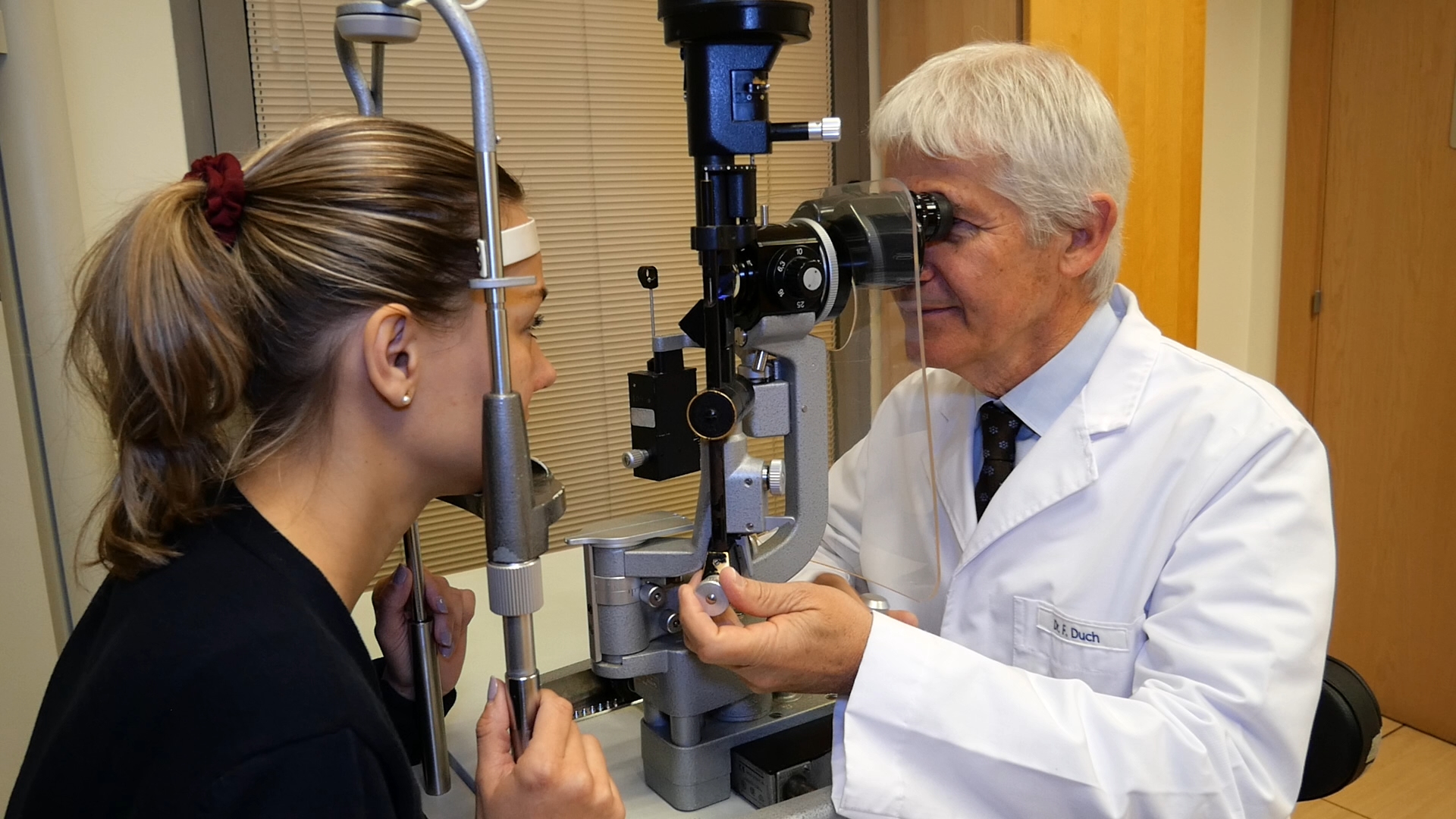
The department in charge of performing the test is the Department of Optometry, which performs all the ophthalmological tests necessary for the diagnosis and follow-up of the different ocular pathologies and the entire visual system. The Department has the most advanced diagnostic equipment and the most modern technology, and its more than 50 optometrists are specifically trained to perform these tests.


Resultats d'una microscòpia especular

Results of a specular microscopy


Frequently Asked Questions

Can it cause any kind of discomfort?

No, since it is a non-invasive technique, it does not contact the eye, so it does not cause any harm.

Do I have to be accompanied?

No, this test does not require you to be accompanied by another person.

Do I have to come fasting?

It is not necessary to come fasting for the realization of the test.

How long does it take to obtain the test results?

The data are obtained at the same time the test is taken. If an ophthalmologist’s report is required, the reported test will be returned within a few days.

Can the optometrist interpret the test result at the time of the test?

The optometrist is the one in charge of performing the test and has the knowledge to confirm its correct performance. The one who has to interpret and report the results obtained is the ophthalmologist, who will do it having the clinical context after a complete anamnesis and examination of the patient.

What are the normal values obtained in a specular microscopy?

The normal values in this test are not exact numbers. They cannot be determined by interpreting the test alone. The ophthalmologist interprets them in the context of the patient’s symptomatology and ophthalmological examination.

Is it possible to drive after the test? And take a shower?

Yes, after the test it is possible to carry out any day-to-day activity, such as driving or taking a shower.


More information

What Specular Microscopy Reveals About Your Patients. Review of cornea and contact lenses

Specular Microscopy. American Academy of Ophthalmology

Related articles

Dr. Rey has published an article with the results and complications of cataract surgery after a pars plana vitrectomy

Dr. Amanda Rey, ophthalmologist at the Department of Retina and Cataract of the ICR, has published an article in the prestigious international scientific journal Clinical Ophthalmology, which studies the visual result and the complications that occur in cataract surgery in patients who have previously undergone a pars plana vitrectomy. What are the objectives of the […]

El Dr. Jürgens i la Dra. Pighin publiquen un cas clínic sobre el maneig d’una extrusió d’ hàptics bilateral en un pacient amb subluxació de LIO

La Dra. María Soledad Pighin juntament amb el Dr.Ignasi Jürgens han publicat un article que exposa el cas d’un pacient que presentava subluxació de LIO, és a dir, un desplaçament d’una lent intraocular, que havia estat implantada feia més de 10 anys.

La Dra. Dyrda publica un article sobre les lents intraoculars en cirurgia de cataractes al Journal of Ophthalmology

La Dra. Agnieszka Dyrda, oftalmòloga del Departament de Retina i Vitri i del Departament de Cataracta de l’ICR, ha publicat recentment un article titulat “Resultats clínics sobre lents intraoculars difractives, refractives, multifocals híbrides i monofocals” a la prestigiosa revista científica Journal of Ophthalmology. En aquest estudi hi han participat altres investigadors de l’ICR: la Dra. […]

El Dr. Duch explica els beneficis de les lents trifocals

El Dr. Francesc Duch, cap del Departament de Cirurgia Refractiva de l’ICR, ens explica com les lents trifocals ens permeten oferir resultats a un grup de pacients que a més dels problemes de visió de lluny i d’aprop, tenen per exemple una graduació d’astigmatisme associada que és difícil de corregir amb altres mètodes. La lent […]

Do you have any questions?

Contact us or request an appointment with one of our specialists.

Visit us at:

ICR Ganduxer Headquarters

C/ Ganduxer,117
08022 Barcelona See map

ICR Pau Alcover

C/ Pau Alcover 67
08017 Barcelona See map

ICR Cerignola

C/ Cerignola, 14
08022 Barcelona See map

ICR Terrassa

C/ Del Nord, 77
08221 Terrassa See map

ICR Service at Clínica Ntra. Sra. del Remei

C/ de l'Escorial,148
08024 Barcelona See map

Clínica Bonanova Surgical Center

Passeig Bonanova, 22
08022 Barcelona See map

Clinica Bonanova Tests and Treatments

C/ Mandri, 63
08022 Barcelona See map

ICR Ganduxer Headquarters

Telephone number +34 93 378 23 19
Emergencies phone number: +34 93 418 72 27

Opening hours:
Monday to Friday 8 a.m. to 9 p.m.
Saturdays 9 a.m. to 2 p.m.

How to get there More information

ICR Pau Alcover

Telephone number (+34) 93 378 23 19

Opening hours:
Monday to Friday from 8 am to 9 pm. Saturday from 8 am to 2 pm.

How to get there More information

ICR Cerignola

Telephone number +34 93 378 23 19

Opening hours:
Monday to Friday 8 a.m. to 9 p.m.

How to get there More information

ICR Terrassa

Telephone number +34 93 736 34 40

Opening hours:
Monday to Friday from 8:30 a.m. to 2 p.m. and 3 p.m. to 8 p.m.

How to get there More information

ICR Service at Clínica Ntra. Sra. del Remei

Telephone number 902 10 10 50

Opening hours:
Monday to Thursday from 3 p.m. to 8 p.m. and Monday mornings from 8 a.m. to 2 p.m.

How to get there More information

Clínica Bonanova Surgical Center

Telephone number 934 34 09 25

Opening hours:
Monday to Friday from 7:30 a.m. to 8:30 p.m.

How to get there More information

Clinica Bonanova Tests and Treatments

Telephone number +34 93 434 27 70

How to get there More information