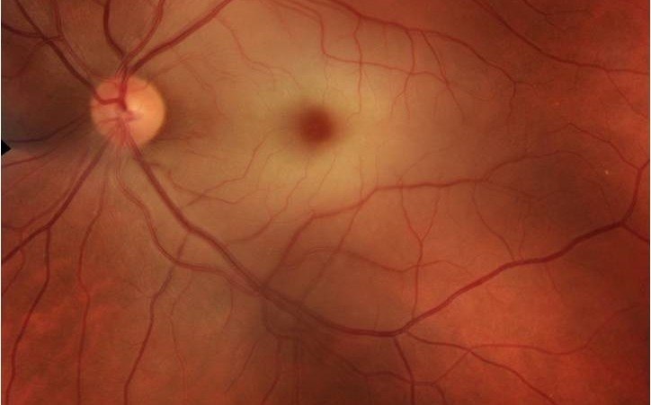
Venous and arterial obstructions in the retina cause a visual loss that can become irreversible and carry greater risks for general health. Diabetes, hypertension, cholesterol and tobacco consumption are risk factors for these cardiovascular accidents.
| Venous occlusions | Retinal occlusions |
|---|---|
| It usually produces a thrombus, whose formation is favored in the course of general diseases (HBP, diabetes, cholesterol …) Causes a retinal whaterlogging It causes a painless loss of vision throughout the visual field or in a part thereof, depending on the affected vein (central or a branch). The organism creates collateral vessels to try to drain the blood, but, in the meantime, intravitreal injections can be administered to keep the retina dry. | It can be produced by the same wall of the vessel, by general diseases that alter it (diabetes, cholesterol …) or an embolism coming from another part of the body. It causes a lack of oxygen in the tissue, and this can suffer a heart attack It causes a painless vision loss and, in the case of emboli, there is a risk that they also reach the brain. In general, there is no effective treatment. Only if it is due to an inflammation of the vessel, it could be treated with anti-inflammatories. It is important to control risk factors to avoid problems in other organs |
As its name suggests, an occlusion of the retinal artery or vein is a blockage in the level of blood flow that runs through this vessel.
Arterial occlusion
Venous occlusion
Thus, an obstructed artery will cause ischemia, that is, a heart attack of the retinal tissue and, in the obstruction of a vein, what will be a waterlogging (oedema) of the retinal tissue.
In vein obstructions, the are usually several causes:
In general, the obstruction of a vein is caused by a thrombus and all the diseases that favor the appearance of thrombi can contribute to the appearance of this vascular accident. Therefore, people who suffer from diseases that affect blood vessels, such as diabetes, hypertension, cholesterol and even those who consume tobacco will be more predisposed.
Only in young patients who have venous obstructions, especially repeated ones, will it be necessary to look further, as diseases related to blood coagulation, that favor thrombotic phenomena.
In arterial obstructions, it must be ruled out that the cause is from the vessel wall itself, as may occur in general diseases such as those mentioned above.
However, sometimes the obstruction is caused by a plunger that comes from another part of the body, usually:
This is important to take into account because when there is an arterial obstruction, we will have to think that there is a risk that the patient not only has an accident at eye level, but also brain.
Another cause of vascular obstructions in both arteries and veins, although less frequent, are inflammations that affect the vessels themselves, which cause:
Patients with obstructions of both arteries and veins will present a painless decrease of visual acuity.
That is to say, they will not feel pain in the eye, nor itching nor stinging, but simply diminution or loss of vision. The loss of vision can be in the entire visual field or in a part of it.
The extent and intensity of vision loss will depend on several factors:
Mainly, the complications involved are visual loss and later, in patients in whom the central vein or artery is obstructed, secondary ocular problems such as glaucoma and, above all, general problems due to their underlying disease can occur; For example, if a source of emboli exists, it should be treated with a cardiovascular specialist to avoid other problems such as cerebral vascular accidents.
To diagnose an artery or vein blockage, simply by performing a deep eye examination (which involves dilating the pupil and looking at the eye with an ophthalmoscope), you can see if there is an artery or vein blockage.
Sometimes, however, to assess the extent of the damage and the monitoring of the treatment, if it can be given, other tests must be done, such as angiography, optical coherence tomography and other types of ophthalmological tests. But simply with an eye fundus revision you can see the cause of the visual loss.
We must take into account, especially in patients with arterial obstructions, that they must be referred to the cardiovascular specialist, because it is very important to rule out that the cause of arterial obstruction is an embolism due to heart or carotid disease, in order to avoid major complications, especially at the cerebral level.
In general, there is no effective treatment, especially if it is a permanent obstruction. Normally, an artery obstruction has a poor prognosis, especially if it affects the central retina, due to the fact that a tissue infarction occurs.
Only in the case that the slowing of the blood stream is due to an inflammation of the vessel, it could be treated with anti-inflammatories.
The organism itself creates a kind of varicose veins that we call collateral veins that try to drain by alternative routes the blood that cannot flow normally through the obstructed vessel. But those collateral vessels, take a while to create.
While the collateral vessels are forming, the retina is waterlogged. Therefore, through intravitreal injections of drugs known as antiangiogenic, or cortisone and, in some cases, the laser, which is used less and less for this pathology, but which is still sometimes necessary, what we do is keep the retina dry so that it does not get damaged while the patient is forming collateral vessels. Later, if the patient has good collateral vessels, the inflammation may disappear on its own, and it may not be necessary to re-treat. However, if the patient does not have good collateral vessels, the retina can occasionally become inflamed and they will have to be treated again so that they do not lose visual acuity.
In general, they cannot be prevented, which is why they are known as cardiovascular accidents.
What is important for prevention is that all patients suffering from general diseases that can affect blood vessels, such as diabetes, high blood pressure, cholesterol, increased blood fat (increased lipids), control these diseases, to avoid, as far as possible, the risk of suffering such accidents.

Contact us or request an appointment with our medical team.