Fluorescein angiography is a test that has been used in ophthalmology for more than 50 years. By injecting a dye into the patient’s vein, we can study the blood flow through the eye to diagnose different retinal illnesses.
There are two types of dye: sodium fluorescein, which was the first to be used, and indocyanine green.
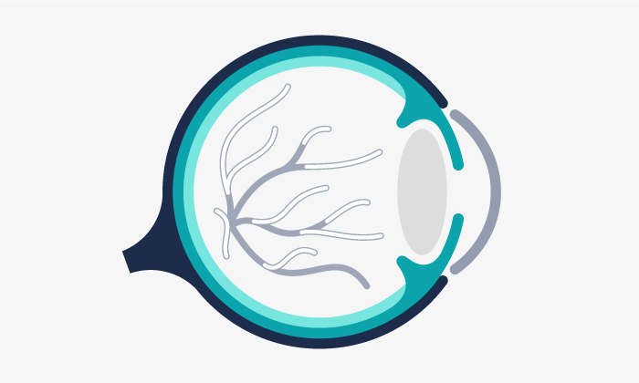
Two dyes are used because they have different properties and uses. There are two types of retinal blood circulation: retinal circulation and choroidal circulation, which also nourishes the retina. Sodium fluorescein is the dye used to study retinal circulation, while indocyanine green is used for choroidal circulation.
The two tests can be carried out simultaneously. First one dye is injected and then the other. Due to the types of filter and the technology that we have we can carry out simultaneous monitoring with both dyes. Fluorescein angiography has many different uses, especially related with vascular pathologies of the retina, such as diabetic retinopathy in its various phases, arterial and vein oclusions and also the study of subretinal membranes in age related macular degeneration, both to classify them and for follow up.
The main contraindication when using these dyes is allergic reactions. For example, if a patient is allergic to sodium fluorescein they should not do this test. Likewise, green indocyanine can cause allergic reactions in those who are allergic to iodine. In addition, we also need to bear in mind cases of acute liver conditions or unmonitored cardiovascular pathologies.
It is advisable for pregnant women to avoid this kind of test as there have been no studies to confirm that it is safe when pregnant.
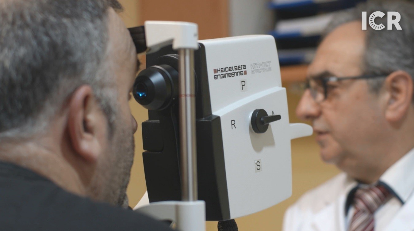
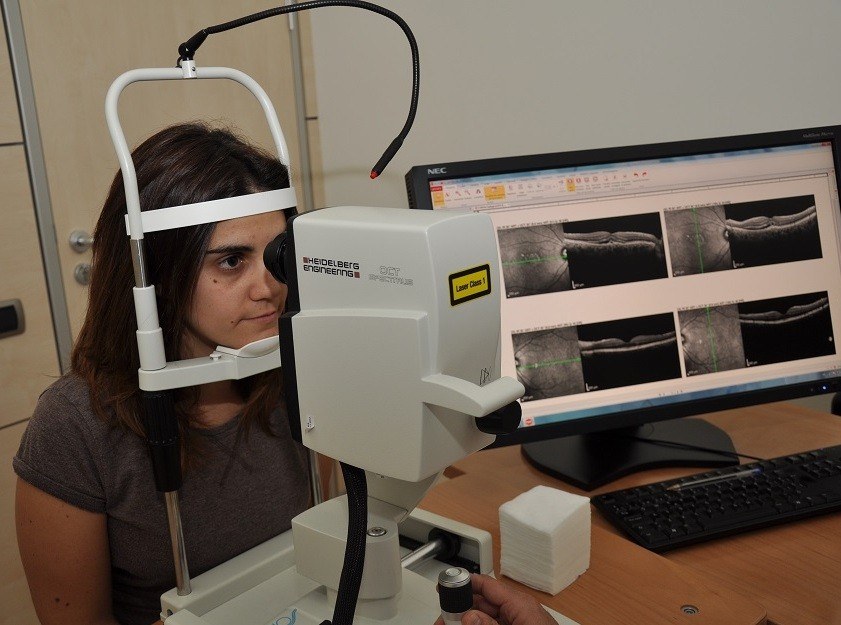
Fluorescein angiography is carried out by using an IV, similar to when you have a blood test. The dye is administered and the passage of the dye through the eye is monitored using a device to achieve images from the retina.
The test lasts for about 10 minutes in the case of the fluoresceinography and, in the case of the indocyanography around 40 minutes. Nevertheless, the total time that the patient spends in the centre from arrival to departure is around 1 hour.
You do not need to fast before the test. You should have breakfast like any other day but not eat excessively.
It is very important to come to the test with someone else, because your vision will be temporarily blurred. Your pupil will be dilated so you will not be able to drive after the test.
The side effects that pupils may experience from the test are the result of pupils being dilated and may include glare and blurred vision. During the test we can see the flashes and lights that are emitted by the apparatus and on occasions we may experience some nausea or temporary heat. In some exceptional occasions stronger sensations may be experienced.
After the test the patient may continue to experience blurred vision for a few hours as a result of the dilating drops. Some patients may also experience a change in skin colour or the colour of their urine, as the dye is eliminated through the urine.
We have a medical and nursing team who are trained in resuscitation and are available at all times in case of an emergency.
The glare effects last for as long as the effects of the eye drops, which can last for around 4 hours, while the change in urine colour can last for 1 to 3 days, depending on the patient’s renal function.
ICR has the best technology on the market. The highly versatile tool that we use can be adapted and focused depending on the patient’s illness to obtain detailed images of the internal layers of the eye, as well as combining the angiography with another test that provides lots of information, the optical coherence tomography (OCT).


AMD it is a pathology that affects the central vision. It progresses as the patient’s age increases and can lead to blindness if not treated early.
Occlusions or obstructions of veins and arteries in the retina are impediments of the blood flow that cause ischemia or flooding. It can cause a painless vision loss to either the entire visual field or only a part of it.
System used to obtain high resolution scan images of the tissues through laser. It is specially useful to study macular diseases and the optic disc.
Contact us or request an appointment with our medical team.


 Request an appointment