
In case of blurred vision, perception of a central spot, straight lines seem bent or you have troubles in focusing when reading, you should go to the ophthalmologist. It could be a central serous chorioretinopaty or other retinal problems. Stress, pregnancy or corticoids intake are some of its risk factors.
| Symptoms of central serous chorioretinopathy |
|---|
| Blurred vision Decrease in vision Central spot in vision Lines seem bent when reading Problems in focusing The objects seem smaller than they are It is very important to go to the ophthalmologist to be correctly diagnosed. |
Central serous chorioretinopathy is not a frequent disease, but it is important to consider it because it is the forth retinal disease that most threatens our vision behind age-related macular degeneration, vascular occlusions or diabetic macular edema.
It is a disease which is usually diagnosed among young male patients, between their thirties or forties, although according to the latest population studies, women may also suffer it. In such cases, it often appears at a more advanced age.
This disease requires a good differential diagnosis, which means it is important to differentiate the disease from other similar diseases, such as polypoidal vasculopathy (a type of age-related macular degeneration), the Vogt-Koyanagi-Harada disease (a type of uveitis), benign tumors (such as nevus or hemangiomas), or more dangerous and malign tumors (such as melanoma or lymphoma).
Patients come to the ophthalmology consulting room reporting:
It is important to differentiate between two types of central serous chorioretinopathy, depending on its length:
Acute central serous chorioretinopathy lasts for a short time and is self-limited (it resolves spontaneously), reason why the visual prognosis is good. In these cases, we need to wait approximately three months in order to assess its progression.
In chronic central serous chorioretinopathy, sequels are more serious. The decrease in visual acuity is caused by anatomical alterations. Irreversible changes occur in the pigment epithelium, a layer located under the retina which is essential to well-functioning photoreceptors. Therefore, it is important to get properly diagnosed in order to determine a treatment, when necessary.
In spite of the high number of studies and interest shown regarding this disease, it is not known for sure. There are several theories. One of the most important ones and with a more solid scientific base is a theory based on the hormonal imbalance, for which glucocorticoids act like mineralocorticoids and cause an inflammation in the choroid, a vascular tissue that nourishes our eyes and is located under the retina.
When there is a vascular inflammation, vessels dilate and become permeable. If there is an increase in blood vessels permeability, fluid leaks out and accumulates on the choroid and is retained by the pigment epithelium. But over time, this barrier becomes inefficient and the fluid runs under the retina. This is how central serous chorioretinopathy occurs.
Besides factors directly related to hormones, other risk factors have been studied:
Diagnosis starts by an identification of symptoms, such as blurred vision, perception of a central spot, bent lines… That is the beginning of it.
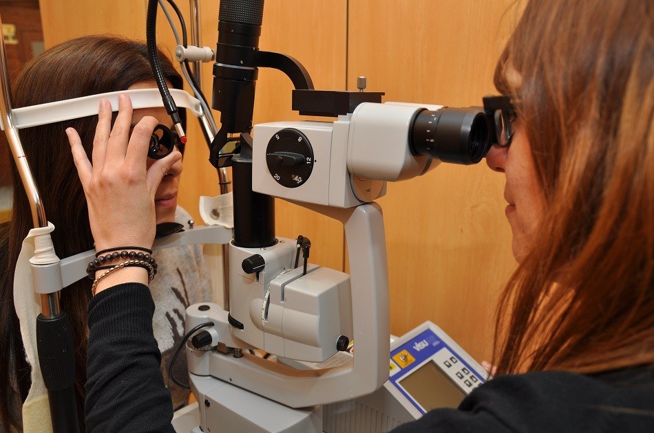
We perform a non-invasive examination, examining the eye fundus with the slit lamp and the use of lenses. We also use a more advanced equipment called OCT (Optical Coherence Tomography). It is a retinal study performed by means of light beams which shows as the retinal tissues and allows us to see in a direct way the liquid accumulated under the retina. It is important the use of the EDI-OCT option, available at our center, in order to examine in detail the choroid, where the disease is located.
In case that the disease is chronic or that we suspect the liquid accumulated under the retina is caused by another disease, we need to use invasive tests empoying a contrast administered intravenously, such as fluorescein or green indocianine angiography. These tests are also used to determine the treatment. They show the state of the choroid and the location of the leakage point through which the fluid accumulated under the retina leaks. There are other functional tests that we may use in order to determine whether photoreceptors have experienced irreversible changes due to the disease. These include visual field, multifocal electroretinogram and microperimetry.
The treatment will depend on the type of central serous chorioretinopathy.
Acute form
In case of an acute central serous chorioretinopathy, it is not usually recommended to apply any treatment, as in 80-90% of cases, it resolves on its own. The only thing we need to do is to monitor the patient and perform the necessary regular controls.
Chronic form
In case the disease does not resolve on its own, is recurrent or the patient has very low vision or requires good vision for professional matters, a treatment must be planned.
Other types of treatments that are currently being studied include the use of micropulse laser. It seems a good alternative, although the study results are still to be confirmed.
Patients are also treated with anti-VEGF intravitreous injections of a substance known as vascular endothelial growth factor, whose use is known as a treatment against ARMD or diabetic macular edema. This drug is currently widely used in ophthalmology.
There are also pharmacological treatments with still non proved scientific based, but the first studies performed reveal its possible efficacy. These are mineralocorticoid inhibitors, such as epleronone or spironolactone.
In case you have symptoms like:
It is important to go to the ophthalmologist not only because it might be a central serous chorioretinopathy, whose chronic form requires in most of the cases a treatent, but also because a good differential diagnosis is needed for an early diagnosis of diseases like ARMD, that has the same symptoms.


AMD it is a pathology that affects the central vision. It progresses as the patient’s age increases and can lead to blindness if not treated early.
Treatment of subretinal neovascularization in age-related macular degeneration. Patients are asked to protect their skin and eyes for 24 hours.

The term uveitis refers to a range of inflammatory conditions that affect the middle layer of the eye known as uvea. Different types of uveitis have different symptoms, causes and treatments.
Contact us or request an appointment with our medical team.


 Request an appointment