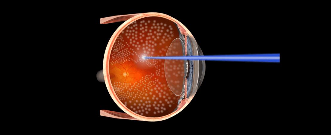
Photocoagulation is a therapeutic technique used in ophthalmology in which a scar is purposely made on the retina using a laser beam. This scar can slow down the progress of certain retinal diseases.
Photocoagulation can stimulate scarring and thus improve the retina’s connection with the wall of the inner eye (for example, in retinal tears and detached retina). It can also improve oxygen supply to certain parts of the retina where this is low, stop unwanted leakage of fluids and help to control development of tumours.
The diseases most commonly treated with this technique are as follows:
To determine whether photocoagulation is the right treatment, a complete eye exam should be carried out. In the case of vascular diseases or tumours the study should be carried out with a fluorescein angiography and an OCT or optical coherence tomography. With these tests the liquid that has accumulated in the suspected area can be seen as can irregular blood vessels and any other defect that could be treated with photocoagulation.
The procedure requires a topical anaesthetic (eye drops). The duration of the treatment may vary depending on the area to undergo photocoagulation and the extent of the injuries.
The laser is carefully focused on a precise area of the retina and fast and repeated laser beams are directed to produce a burn only in the desired zone, avoiding neighbouring areas. When the zone to be treated is more extensive, what is known as panretinal photocoagulation or panphotocoagulation can also be carried out.
The procedure is carried out under topical anaesthesia and is not normally painful. In cases of extensive photocoagulation such as panphotocoagulation, anti-inflammatory eyedrops can be prescribed. In extreme cases, depending on the patient’s pain threshold, injectable anaesthetic should be used to prevent pain.
Despite the various studies that demonstrate the effectiveness of this treatment, in some cases photocoagulation can be ineffective or can need to be complemented with other procedures.
In addition, we must remember that all procedures have some risk. In the case of photocoagulation there is a risk that the laser beam will cause some kind of haemorrhage or excessive scarring that damages eye structures such as the macula.
The glare effect caused by the laser beam may last for some time. After treatment, vision may be slightly reduced and then recover two to six weeks later. Provided that the retina was not previously damaged, vision may subsequently improve. This will depend on the problem being treated, the eye’s response to the treatment, and the degree to which the eye was affected. In the case of retinal damage, laser may slow down the rate at which the disease progresses.


In most cases, the vision can be recovered after a surgery for retinal detachment: But if it is not treated, the patient’s vision could be irrevocably lost.
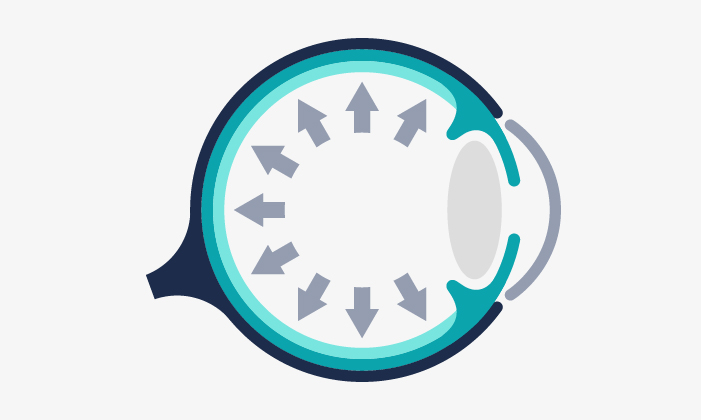
Glaucoma entails a loss of the vision field, usually asymptomatic until later stages of the disease. If it is not treated properly, it can induce blindness.
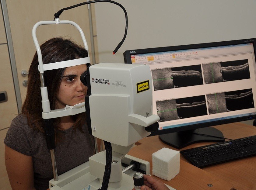
System used to obtain high resolution scan images of the tissues through laser. It is specially useful to study macular diseases and the optic disc.
Contact us or request an appointment with our medical team.


 Request an appointment