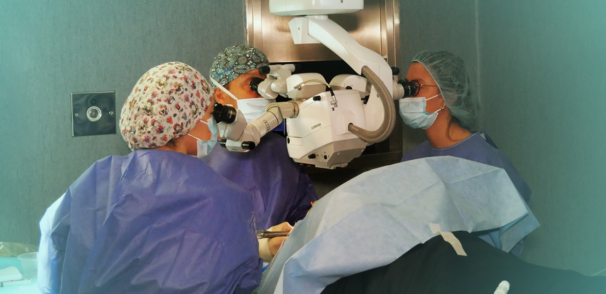
Scleral surgery is a surgical procedure that is normally carried out under local anaesthesia (or general anaesthesia in specific cases). It involves placing a ring, usually made of silicone, around the most external layer of the eye wall, the sclera. This ring remains sutured to the wall of the eye ball, over the area where the retinal tears are found.
The ring pushes the sclera inwards by exerting pressure. In this way it relieves the tension that is pulling on the retina and controls the traction that causes the retinal tears. It can be sutured to apply pressure to the area behind the tear or it can encircle the eyeball 360º.
Releasing the traction alone is not enough to block the tear which is why it needs to be combined with a procedure that causes inflammation and then scarring of the tear. This process is achieved by applying cold (cryocoagulation) or light (photocoagulation with laser). Scars are thus formed on the retina which are sealed with layers below and help to hold it in place.
Sometimes the fluid beneath the detached retina needs to be drained. In doing this we enable the retina to be reposition itself against the back wall of the eye.
In practically all cases of detached retina, the retina needs to be repositioned to avoid permanent loss of function which could lead to blindness.
Simple scleral surgery is useful for treating retinal tears or holes that have led to a detachment which is not overly complicated. The procedure should be carried out urgently as, in the most acute cases, the longer it takes to repair the detachment, the less possibility to correctly reattach the retina without the need for more complicated techniques with a greater risk of affecting the final visual outcome. For this reason, it is normally necessary to reapply the retina within a couple of days.
As well as requesting your complete medical history, we need to carry out a full eye exam to determine the area that needs to be treated by surgery. For this a slit lamp exam of the anterior segment (cornea, lens…) and posterior segment (macula, retina…) of the eye is carried out and sometimes other tests such as ocular ecography or OCT.
After surgery, patients should follow the postoperatorative instructions from the surgeon. These may include not lifting heavy things, resting and not doing any strenuous activity.
Rapid eye movement should also be avoided and patients should wear sunglasses outside. Your eyes might be red and you may have the sensation that you have something in your eye or a slight discomfort for a few days after surgery. The ophthalmologist will prescribe antibiotic drops and corticosteroids to reduce the swelling and the risk of infection.
In cases of excessive pain, swelling, bleeding, secretions or reduced vision, you should inform your ophthalmologist as soon as possible.
Until 6-8 weeks after the surgery, your best visual acuity cannot be determined. Complete visual recovery will depend on the location and seriousness of the detachment.


In most cases, the vision can be recovered after a surgery for retinal detachment: But if it is not treated, the patient’s vision could be irrevocably lost.

A macular hole causes loss of central vision and distortion in the images and is manifested with undulation of straight lines. There are several surgical procedures that can be performed to treat it.

A vitreous detachment is the separation of the vitreous humour from its attachment points on the retina. This causes a sudden appearance of spots in the form of spider webs or flashing lights.
Contact us or request an appointment with our medical team.


 Request an appointment