Vitrectomy is a type of eye surgery to treat problems of the retina and the vitreous. Retina is a light-sensitive tissue located in the back of the eye. Vitreous is the clear gel substance that fills the eye.
Vitrectomy consists of the extraction of the vitreous humor fluid and, usually, replacing it with a saline solution or with a bubble of gas, air or silicone oil. When the vitreous is replaced by gas or by silicone, it may be necessary for the patient to lay face down or on his side for a few days during postrecovery. Moreover, if the inside of the eye is filled with gas or air, it is forbidden to fly in an airplane or travel up higher than 500 metres above the sea level until the gas bubble is gone, since rapid altitude changes can affect it.
The ophthalmologist may consider necessary to perform a vitrectomy when one of the following eye problems is present:
Vitrectomy can often improve or stabilize vision. The procedure removes bleeding or the remains of an infection or inflammation that could block or blur the light by focusing on the retina. Moreover, it also eliminates scar tissue that could move, tear or rip the retina.
This surgery can also help to remove a foreign object, that has remained inside the eye after a perforating trauma,and that could damage vision if not removed.
The ophthalmologist may recommend performing an ocular ultrasound scan, Retinal Optical Coherence Tomography, or other texts to examine the eye.
Vitrectomy is usually performed in an outpatient surgery center and it takes between half an hour to several hours. It is performed under local or general anesthesia to numb the eye.
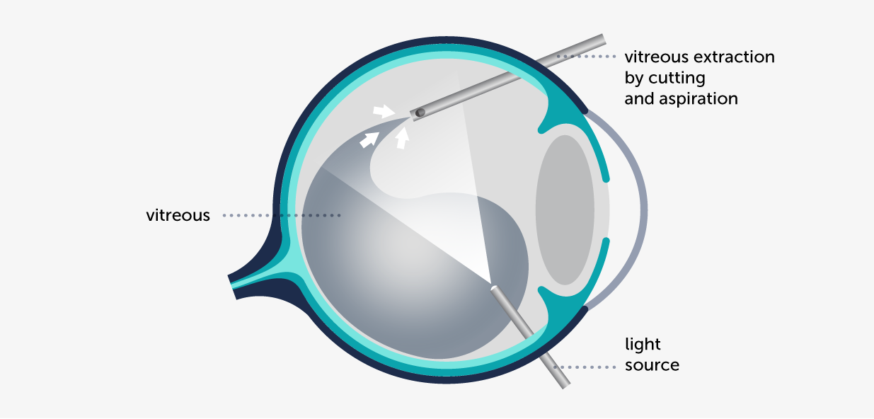
During surgery, the ophthalmologist makes three small cuts or incisions of less than 1 millimeter in the white of the eye, the sclera, with the use of a microscope to see inside the eye. The surgeon will use tiny surgical instruments to do one or more of the following steps:
The ophthalmologist will prescribe medicine to relieve pain and eye drops that should be used for several weeks. The ophthalmologist will also tell the patient when to go back to normal activities.
Like any other surgery, vitrectomy has risks. Nevertheless, they are by far outweighted by the benefits of improved vision.
Some of the riscks are: bleeding, detached retina, high intraocular pressure or infections.


There are different kinds of eye injuries, caused by blunt or perforating objects as well as chemical products. If not treated properly, they could have serious consequences.

Macular epiretinal membrane causes a slow and progressive visual loss lasting months or years, and affecting one or both eyes. Generally, it causes image distortion, and undulation of straight lines.

A macular hole causes loss of central vision and distortion in the images and is manifested with undulation of straight lines. There are several surgical procedures that can be performed to treat it.
Contact us or request an appointment with our medical team.


 Request an appointment