
Dr. Amanda Rey, member of the ICR Cataract, and Retina and Cornea Department, has published in the journal Annals d’Oftalmologia an article about Macular Retinoschisis, a common pathology in patients with high myopia.
Macular retinoschisis or foveoschisis is an eye pathology produced by the separation of the layers of the central area of the retina (macula) and can cause distortion of straight lines (metamorphopsia) and loss of visual acuity. According to established protocols, surgical intervention is only recommended in those cases where there is visual worsening secondary to disease progression.
Within this disease, there are different clinical forms:
It is also classified according to the traction exerted on the macula, which can also cause this disease. The traction may come from the vitreous (anteroposterior traction) or from an abnormal tissue that forms over the macula, the epiretinal membrane (tangential traction).
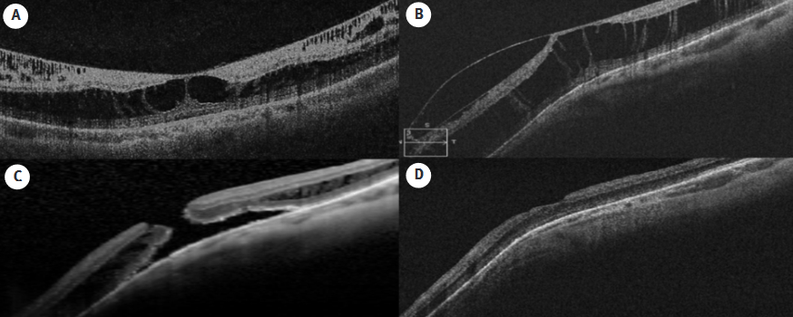
For its diagnosis, a non-invasive test called optical coherence tomography (OCT) is needed, which allows to classify the pathology and to carry out the appropriate treatment.
Dr. Rey had previously published an article on myopic foveoschisis in the prestigious international journal Ophthalmologica, where she analyzed 56 eyes of patients from the Institut Català de Retina with myopic foveoschisis and classified them by OCT.
This article studied the follow-up of these patients and observed that 71% of the cases remained stable and only 29% required surgery in the first year of follow-up. It also analyzed the evolution of the patients according to the type of retinoschisis and the treatment used in each case:
The results obtained in this study are positive, since the protocol established by our center achieved anatomical and functional recovery in 75% of the cases operated on 3 months after surgery and 81% of the patients had gained vision.
In short, we see that good ophthalmologic control is essential to facilitate early diagnosis and treatment of ocular diseases related to high myopia, including macular retinoschisis.
The study also involved Dr. Ignasi Jürgens, ICR Medical Director, la Dr. Agnieszka Dyrda and Dr. Xavier Maseras, members of the ICR Retina and Vitreous Department.


Contact us or request an appointment with our medical team.


 Request an appointment