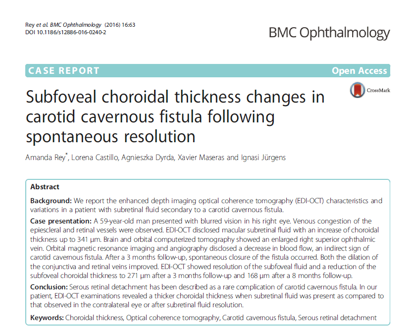
Dr. Amanda Rey, ophthalmologist of the Retina Department at Institut Català de Retina, alongside Dr. Lorena Castillo, ophthalmologist at ICR Neuro-Ophthalmology Department, have published an article in the prestigious international ophthalmology journal BMC Ophthalmology.
The featured article reports a clinical case of a patient diagnosed with carotid-cavernous fistula with loss of vision caused by a secondary serous retinal detachment.
A carotid-cavernous fistula is an abnormal connection between the carotid arterial system (which carries oxygenated blood to the brain) and the cavernous sinus which can typically cause a venous congestion in the eye. Serous retinal detachment has been described as a very infrequent complication in patients suffering from carotid-cavernous fistula.
Choroid is a vascular layer that nourishes the external retina and is involved in several ocular diseases, such as tumours, macular degeneration, central serous chorioretinopathy, diabetic retinopathy and uveitis. EDI OCT (Enhanced Depth Imaging Optical Coherence Tomography) is a non-invasive technique allowing to evaluate the thickness of such layer, as well as these diseases’ structure.
The analysed results for the patient confirm higher subfoveal choroidal thicknesses at the time of fistula diagnosis with subretinal fluid presence when compared to the other eye or to the thicknesses observed after the fluid resolution, when the fistula has spontaneously closed. Therefore, this is a disease in which the origin or cause of the occurrence of secondary serous retinal detachment could have something to do with the choroid.
It is speculated that the transformation of venous blood of orbital veins into arterial blood due to the carotid-cavernous fistula could cause venous stasis (slow blood flow in veins), congestion of capillary lamina of the choroid and, finally, it could lead to an hypoxia (state of oxygen deficiency in blood, cells and tissues), thus causing a dysfunction of retinal pigment epithelium cells, which leads to a secondary serous retinal detachment. Changes in choriocapillaris congestion can be measured by choroidal thicknesses in a non-invasive way using an EDI-OCT. When the carotid-venous fistula is closed, the venous flow in the eye area goes back to normal, and so does the choriocapillaris. In the same way, the pigment epithelium becomes normal as well, the serous retinal detachment is resolved and the normal choroidal thickness is restored.
This is the first published paper revolving on a patient with carotid-venous fistula and secondary serous retinal detachment in which the EDI-OCT technique has allowed to analyse the changes experienced by the choroid after the spontaneous resolution of the fistula. This technique allows to visualize in real time and in an objective and detailed way the choroidal layer and it can also be used to monitor the disease’s activity.
Dr. Ignasi Jürgens, ICR Medical Director and Head of Retina Department, as well as Dr. Agnieszka Dyrda and Dr. Xavier Maseras, both ophthalmologists at ICR Retina Department also collaborated in this paper.

Contact us or request an appointment with our medical team.